Pathological findings within the group of animals
PMWS is a disease of the lymphoid tissues with secondary lesions in the other major organs. Note PMWS cannot be diagnosed on postmortem findings only as these findings can be recognized on many farms without the clinical signs of PMWS.
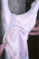
The lymphoid tissues in the acute phase presents as enlargement and are more noticeable/prominent lymph nodes – often in areas where lymph nodes would not be noticed. Three areas of lymphoid enlargement should be examined:
The superficial inguinal lymph node
The abdominal lymph nodes – mesenteric (which can be extremely variable), hepatic and gastrosplenic.
The bronchial lymph nodes
|
|
|
|
|
Superficial inguinal lymph nodeThese may become more bilaterally prominent and may even be visible to the naked eye |
|||
|
|
|
|
|
|
Mesenteric |
Hepatic |
Gastric |
|
|
Abdominal lymph nodes – in particular the mesenteric, hepatic and gastrosplenic lymph nodes demonstrate enlargement – this can be extremely variable. |
|||
|
|
Bronchial and chest lymph nodes are enlarged When
there are respiratory problems the bronchial lymph nodes can become
prominent. The ones shown are only
slightly enlarged. |
||


Other gross pathological findings which are commonly associated with PMWS
|
|
|
|
Pericardial effusion – fluid around the heart |
Rounding of the heart – enlarged right venticle – this may be the cause of the fluid around the heart and other organs |
|
|
|
|
Pale enlarged liver – this may be associated with jaundice or icterus - a yellowing of the carcass |
Gastric ulceration |
|
|
|
|
Enlarged kidney |
Kidney with “white spots” which are caused by nonpurulent interstitial nephritis. |


With secondary infections becoming involved, the pig also presents with the range of pathology associated with each of these secondary infections which are then superimposed over the lesions described above.
Laboratory findings
Histological examination of the enlarged lymph nodes reveal a characteristic
lesion. Note however, this lesion can
also be found in wasted/compromised pigs without PMWS, especially when only single
lymph node is examined. It is essential
that at least tissues samples from at least 3 different enlarged lymph nodes
are presented from each affected animal to demonstrate that the condition is
systemic.
|
|
|
|
|
Normal lymph node Each of the follicles (one
highlighted by the circle) shown are a single B lymphocyte clone line of
cells producing a single antibody |
PMWS lymph node In the infected lymph node
the B cells have disappeared from the follicle and only the background
remains – highlighted by the arrow |
Overwhelming PCVII in tissues – example lung The brown areas are areas
of PCVII virus |
|
|
||


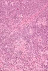

The depleted follicle is replaced with large histiocytic cells and giant multinucleate cells. Cytoplasmic PCVII viral inclusion bodies may be seen in the histocytes or dendritic cells. Note with time the lymph nodes may actually undergo atrophy (a reduction in size).
|
|
|
|
Multinucleated cells in the lymph node |


When the lymph node is examined for the presence of PCV II in positive cases of PMWS there will be an overwhelming presence of PCV II in the macrophages of the lymph node.
|
|
|
|
|
|
|
|
|
|
||