Enzootic
(Mycoplasma) Pneumonia
|
Also called |
Virus pneumonia.
Mycoplasma pneumonia. EP. PRDC – Porcine Respiratory Disease Complex. |
||||
|
Causal
agent |
Mycoplasma
hyopneumoniae. A mycoplasma does not have a cell wall |
||||
|
Occurrence |
A disease
commonly seen in growing and finishing pigs Note enzootic
pneumonia may not require Mycoplasma
hyopneumoniae – it just describes the clinical condition |
||||
|
Complicating
factors in the clinical expression of enzootic pneumonia |
|||||
|
Bacteria |
There are a
number of bacteria and mycoplasma which can infect the lung, particularly
after the effects of the mycoplasma on the mucocilary escalator. These include pasteurella, streptococci and
Actinobacillus pleuropneumoniae
(APP), Actinobacillus suis, Haemophilus parasuis (Glässers
disease) also plays a contributing role in post-weaning respiratory disease. |
||||
|
Viruses |
These include PRRSv,
Swine Influenza, Circovirus II and Porcine Respiratory Coronavirus (PRCV).
Aujeszky’s Disease (Pseudorabies) and Classical Swine Fever may play a
pivotal role. |
||||
|
Others |
Parasites-
Ascaris, lungworm |
||||
|
Environmental
factors |
There are many
factors for which the stockperson is responsible. These include: |
||||
|
Air |
Excessive 24
hours temperature variations. Draughts. High ammonia concentrations |
||||
|
Floor |
Overstocking. Rough floors |
||||
|
Water |
Poor water flow.
Insufficient drinkers |
||||
|
Feed |
Dusty feed. Poor feed availability |
||||
|
Clinical signs |
Coughing, with or without fever (with fever 40.5 to 41C implies a
complicated enzootic pneumonia), laboured
breathing, variable growth rates, unthrifty appearance, reduced appetite and
increased post-weaning mortality. Classically
the clinical signs are seen in pigs of 60-80 kg, but in complicated cases can
occur much earlier. Note the piglet
may be infected from 14 days of age without any clinical signs. If in naïve herds
break, sows may abortion due to pyrexia. |
||||
|
Transmission |
The disease can
move via the air from infected farms to adjacent farms within 3 km. On infected
farms, the disease is transferred from the sow/gilt to her offspring, sows
may still have mycoplasma in her nose at parity 8. Infected pigs spread the disease by droplet spread from nose to nose contact and coughing pigs. One cough can spread the disease 4 metres, assuming the
mycoplasma can survive the cough |
||||
|
Incubation |
With high level
of infection, incubation takes 5 days.
With a moderate level of infection incubation may take 4 to 6 weeks |
||||
|
Effects
of enzootic pneumonia |
Depending on the
extent, enzootic pneumonia can reduce
daily liveweight gain by 17% and increase feed conversion by 14%. In other cases causes death. Enzootic pneumonia can also have a
significant effect on PRRSv infections making them more serious to the weaner
by encouraging macrophages into the lung. |
||||
Pathogenesis |
Mycoplasma hyopneumoniae
graze the cilia
on the trachea and bronchi (the windpipe).
The cilia are important as they help to protect the lung from
particles (dust and disease). Once the
disease enters the lung it causes areas to
collapse and the pig progressively becomes
short of air. The collapsed areas
become infected with other diseases and the pig finally succumbs to the
disease load. The mycoplasma has an
effect on macrophages and reduces their ability to kill and digest other
pathogens. There is a significant
effect of coinfection risk with PRRSv and Aujeszky’s disease which will
potentate the clinical signs |
|
|||
|
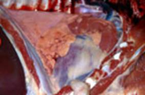
|
The
diseased collapsed areas are darker (arrow) than the normal light parts of
the lung |
The normal lung floats
while the diseased lung sinks |
|
||
|
Diagnosis |
Slaughterhouse
examination |
|
|||
|
Examination of
the serum by ELISA and PCR. Immunohistochemistry
of tissue sections |
|
||||
|
Treatment
and control |
Greater than 70% of normal health herds are infected by Mycoplasma hyopneumoniae. Because the disease is so widespread, control and
treatment is complicated. Elimination
of Mycoplasma
hyopneumoniae – specific
programmes |
|
|||
|
EP
- ve herds |
Where herds are set up from EP-ve pigs, these herds have much
less problems with respiratory disease.
Maintenance of the EP status takes a lot of time and planning. Sitting of such a pig farm is fraught with
difficulty as the mycoplasma can spread 3km through the air. Note the proximity of major roads. On negative units, it still may be worth vaccinating gilts
to protect the adult herd from an abortion storm and pig flow disruption. |
|
|||
|
EP
+ve herds |
|
|
|||
|
Eradication |
Difficult both practically and economically as herds can
be re-infected quickly. May even be
impossible on certain units.
Tulathromycin may be used in an elimination programme Elimination of Mycoplasma
hyopneumoniae – specific
programmes |
|
|||
|
Antibiotics |
Antibiotics limit the effects of the disease. However,
subsequently to viruses becoming involved in pig respiratory disease,
antibiotics are proving less effective |
|
|||
|
Herd
management |
Improvements in the environment of the pig greatly help to
reduce the stress factors. In particular improvements in ventilation
and a reduction in the stocking density should be attempted |
|
|||
|
Disease
management |
Partial depopulation, cleaning and repair of the
growing/finishing phase has helped considerably. This may be combined with all-in/all-out,
effective pig flow, batching
systems and 2 or 3
site production systems |
|
|||
|
Vaccination |
Mycoplasma hyopneumoniae vaccines significantly
help to reduce the effect of the disease.
The vaccine is administered between 7 to 10 days of age and at weaning
(21-28 days), but awareness need to be made regarding the maternal antibody
levels provided from the sow Do not
vaccinate the sow to raise maternal antibodies. Vaccinate gilts and boars as part of their introduction
period. |
|
|||
|
Zoonotic Implications |
|
||||
|
|
None |
|
|||

Enzootic
(Mycoplasma) Pneumonia
The
approximate relationship between lung damage/scoring system
at
slaughter at 95 kg and daily liveweight gain and food conversion ratio
|
|
Lung
Lesion |
DLW
Reduction |
FCR
increase |
||
|
% |
gr./day |
% |
Value |
||
|
|
0/55 NEGATIVE |
0 |
0 |
0 |
0 |
|
|
2/55 MILD |
-4 |
-25
|
0 |
0 |
|
|
10/55 MILD |
-7 |
-50
|
+5 |
0.15 |
|
|
15/55 MODERATE |
-11 |
-80
|
+8 |
0.25 |
|
|
20/55 MODERATE |
-15 |
-100
|
+11 |
0.35 |
|
|
30/55 SEVERE |
-20 |
-130
|
+14 |
0.40 |
|
|
55/55 SEVERE |
-22 |
-560
|
+17 |
0.50 |


The estimates of reduction in DLW and FCR is based on Straw 1989 using pigs with 700g/day over the finishing period and a FCR of 3.
The lungs are shown from the
ventral, with the intermediate lobe superimposed for completeness
The
severity of the lesion may indicate stage of infection. Therefore, an individual pig with a severe
lesion may have been only recently affected and have excellent growth
rates. This can be compared with the
growth rate of chronically affected (smaller lesion) pigs.
Specific Mycoplasma hyopneumoniae
elimination programmes