Anatomy of the honey bee – Apis mellifera
The honey bee -
Class – insect
Order –
Hymenoptera
Family – Apidae
Genus – Apis
Species - mellifera
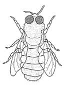
Honey bee castes
|
|
|
|
|
Queen |
Worker |
Drone |

Note the QUEEN is large with a long
tapered abdomen and the wings are shorter than the body. The queen lays fertilized eggs – workers and
unfertilized eggs – drones.
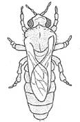
The DRONE is a male they have no
sting. The eyes cover most of their head
and the wings are as long as their bodies.
They have a blunt tipped stocky abdomen.
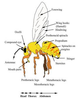
External anatomy
|
|
The figure illustrates the basic surface anatomy of
the honey bee – Apis mellifera. Note the size difference and slight shape differences
between the queen, worker and drone.
Especially note the large eyes of the drone – useful in the mating
flight. |
Internal general
anatomy of a honey bee
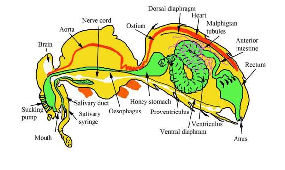
Photograph of the
digestive tract of the bee (with the head end removed)

Pheromones glands
The bee contains several glands.
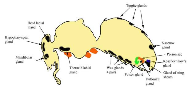
Nasanov gland
This produces a variety of chemicals which the bee uses
to assist identification of the entrance of the hive.
Koschevnikov
gland
This releases alarm pheromone – attracts other bees to
attack and sting the same part of the body of the offending animal. Several compounds – principle one is
isopentyl acetate.
In the queen this gland products are responsible for
the formation of the clusters of court bees that surround the queen.
Dufour’s gland
The products of this gland line the entrance to the
hive and may assist recognition of family or nest ownership
Mandibular glands
In young workers this gland produces the lipid-rich
white substance mixed with the hyopharyngeal gland secretions resulting in
royal jelly.
In older worker this produces part of the alarm
pheromone.
In the queen, this gland has a number of important
functions – produces queen substance (queen mandibular substance) and is
associated with:
Suppression of construction of emergency queen cells
Inhibits ovary development in the workers
Attracting drones during the mating flight
Attracts the attendant workers
In the drone, the mandibular gland assists in the
formation of drone gatherings – in drone congregation areas (DCA’s) which
appear in open fields.
Hypopharyngeal
glands
Produce protein-rich sections (Royal jelly) when the
worker is a nurse bee.
When the worker becomes a forage bee it produces
invertase which helps break down sucrose into fructose and glucose.
Pre-tarsus gland
As yet its function is not known.
Arnhart or
footprint glands on each foot
The reproductive organs
Worker
reproductive organs
|
|
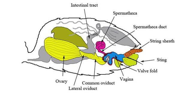
The female worker reproductive tract Which in the worker develops in the stinging
gland. Note the stinger has two
components, the poison gland (filled with colourless liquid when fresh) and
the alkaline gland (which may appear yellow) together with the stinger. Each sting contains 150 mg of venom. Note bees, unlike wasps, die after using the stinger
as the organ is left in the victim.
|
||
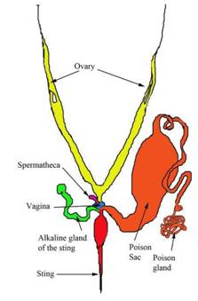
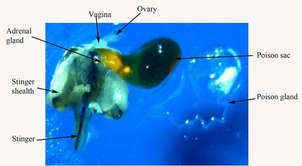
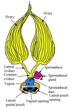
Queen
reproductive organs |
|||
|
The queen reproductive tract. A single ovariole |
During the mating flight the queen the drone’s penis
bulb is discharged by eversion of the penis into the queen’s vaginal pouch. After mating the queen separates from the drone and
the male penis bulb remains in the female. The queen may mate with several drones during the
flight. The spermatozoa are discharged in the distended
lateral oviducts. Once back at the hive, the workers remove the penis
bulb from the queen. The spermatozoa
are then moved into the vagina and then the spermatheca gland where they
remain for the productive life of the queen – up to 5 years. If the queen “runs out of semen” she will only lay
unfertilized drone eggs. |
||

The abdomen on
the queen bee – concentrating on the reproductive organs
|
|
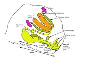
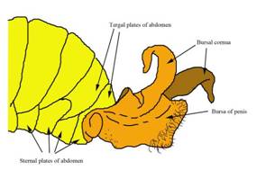
Male
reproductive tract
|
|
During mating, sperm mass stored in the bulb of penis
is discharged by eversion into the queen’s vagina. After mating the queen separates from the male with
the bulb of penis remaining in her genital tract. The male reproductive organs tear at the penis
neck. The drone subsequently bleeds to
death. |
|
The male
abdomen with the penis everted |
End of the male
abdomen, ventral aspect |
|
|
|

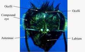
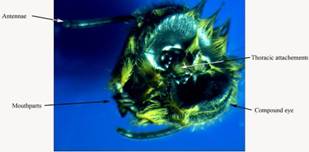
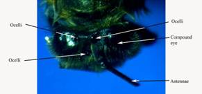
Head of the honey bee – note all activities in the hive
are in complete darkness
All communication
is done by touch, feel and smell
|
|
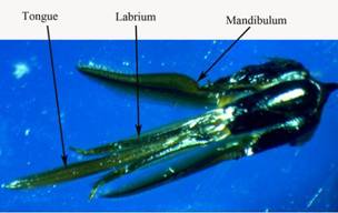
The mouth parts are combined so they can chew and
suck having both proboscis and mandible. Many insects have evolved to possess
only one or the other capabilities.
|


|
|
|
||||||||||||||||||
|
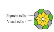
Eyes
Occasionally drones will be born with white
eyes. This is a genetic defect.
Replace the queen. |
||||||||||||||||||


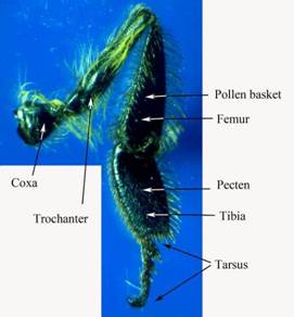
Thorax of the honey bee
|
|
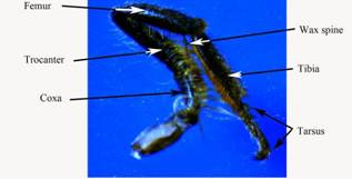
Unlike other insects the bee’s thorax is divided into
four segments – prothorax, mesothorax, metathorax and propodeum. The prothorax carry the prothoracic legs. The mesothorax and metathorax also carry legs and
each of the wings. The claws on the last tarsomere allows bees to walk
on rough surfaces (tree trunks) together with a soft pad (arolium) allowing
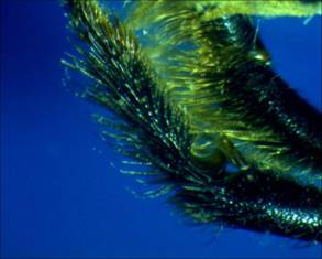
them to walk on smooth surfaces (leaves) The hind legs in the worker are fringed with long,
curved hairs and the space thus enclosed is called the pollen basket or
corbicula. |

|
|
|
|
Prothoracic leg Lateral view |
Prothoracic leg medial view |
|
|
|
|
Detail of the prothoracic antennae cleaner |
Detail of the mesothoracic wax spine |
|
|
|
|
Mesothoracic lateral view |
Mesothoracic medial view |
|
|
|
|
Metathoracic lateral view |
Metathoracic medical view |
|
|
|


Tarsus of the mesothoracic leg
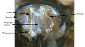
Tracheal system in the thorax
|
|
|
|
The gross appearance of the tracheal system in the
thorax |
With the prothorax tergite removed |
|
|
|
|
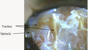
Detail of the main tracheal duct |
Examination of the normal trachea by the microscope |


The development of the wing is a fascinating story of
insect evolution. One possibility are that their origins start as gills.
|
|
|
|
|
|
|
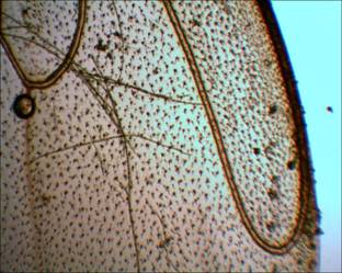
Detail of the forewing limb |
Detail of the hamuli – wing hooks |